

In brief
- A Copenhagen team combined PET and MRI scans to observe, simultaneously in the same brain, how LSD affects 5-HT2A receptors and blood flow.
- LSD occupies these receptors with high affinity, increases global cerebral blood flow, and reduces global connectivity, particularly in visual and attention networks.
- The complete data set includes only seven volunteers, making this a mechanistic study rather than a clinical efficacy trial.
It has been known for decades that LSD acts on the 5-HT2A serotonin receptor, but measuring this in a living person, with known doses, while simultaneously recording brain activity, is significantly more challenging. A study published October 12 in Nature Neuroscience attempted this using an uncommon technique: simultaneous PET and functional MRI.
A dual scan and blinded doses
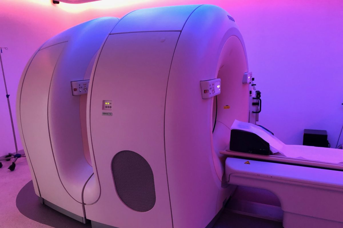
The work comes from the Neurobiology Research Unit at Rigshospitalet in Copenhagen, in collaboration with the University of Copenhagen and the University Hospital of Basel. The healthy volunteers took oral LSD in doses ranging from 25 to 200 micrograms without knowing which dose they received. Inside the scanner, a radioactive tracer ([11C]Cimbi-36) allowed researchers to see how much of the 5-HT2A receptor was occupied, while MRI sequences measured cerebral blood flow, flow in the internal carotid artery, and connectivity between regions.
The final sample size is small. Only seven volunteers provided complete data at the time of peak effect. Four did not complete the LSD scans: two due to equipment failure and two due to adverse effects. As a reference, the authors used another group of 25 people who had received psilocybin.
What was observed in the brain
According to the article’s abstract, LSD binds to the 5-HT2A receptor with high affinity, and there is a relationship between dose and receptor occupancy that, in the authors’ opinion, could guide the clinical development of these substances. Additionally, global cerebral blood flow and internal carotid artery flow increased; both measures moved in tandem. Global connectivity decreased, especially in visual and attention networks in most participants, and this decrease was inversely associated with the increase in blood flow.
One pharmacological detail stands out: when crossing plasma LSD concentration with subjective intensity, the curve draws a “counterclockwise” loop. Simply put, the effects do not follow blood levels point-for-point but rather move at their own pace. The authors interpret this as atypical pharmacodynamics. With this sample size, it is best to view this as a clue rather than a definitive conclusion.
What it implies
The value of the study lies in the method: being able to quantify the relationship between dose, receptor, and vascular response helps explain why LSD lasts so long and why two people taking the same dose may have different experiences. It also serves as a reminder that part of the signal measured with MRI reflects changes in blood flow, not just neuronal activity—something to keep in mind when interpreting other imaging studies.
The limitations are clear. Seven healthy people, variable doses, and a single laboratory setting do not allow for extrapolation to therapy or use outside the lab. It aligns with other recent work on the rhythm of cortical activity under LSD, such as the one we previously reviewed. To place the substance in its historical and pharmacological context, you can consult our guide on LSD and ergot.
Source
- McCulloch, D. E.-W., Larsen, K., Fisher, P. M., Knudsen, G. M. et al. Investigating molecular and hemodynamic effects of LSD in the human brain. Nature Neuroscience, October 12, 2026.
Educational content written from a harm reduction perspective and with respect for individual freedom. It is not a substitute for advice from a healthcare professional and is not intended to encourage or condemn any drug use.